It’s one of life’s little ironies. The proteins in our bodies fight infection, carry messages, ferry oxygen and build tissue. But then, like double agents in a spy novel, they can betray us. They overreact to a virus and attack our own organs. They promote cancer, help clog arteries or set up roadblocks in the brain. We may never know until symptoms appear — a lump, chest pain, severe memory lapses — and irreversible damage is done.
Who wouldn’t like to get ahead of these heart-stopping scenarios? By detecting proteins gone awry or elevated in the earliest stages of disease, scientists are opening up the possibility of effective therapy before health is compromised. The standard checklist on the annual physical — temperature, blood pressure, reflexes, lung function, skin condition — is already backed up by blood tests for molecular markers such as cholesterol and other proteins. What researchers envision is an inexpensive (some aim for a “penny per protein”), accurate and rapid test that can be performed in a doctor’s office and provide unprecedented views of our biochemistry on the fly. In medical research labs across the country, a race is on to identify new biomarkers and to develop highly sensitive technologies that can measure them. Talk about a needle in a haystack. A single blood sample can contain roughly 9,000 types of proteins, although different analytical techniques produce widely varying estimates. And on top of the sheer flood of proteins is the fact that they are shape shifters. Once released into the bloodstream, they may be altered before they reach their intended destination.
Oregon State University chemist Vince Remcho likens the search for proteins to fishing. “Ultimately you are searching for one particular protein or other molecule in a vast soup of molecules, so you have to choose the right bait. My group is in the bait business, bait and hook,” he says.
In Remcho’s lab in OSU’s new Linus Pauling Science Center, some of that bait consists of short relatives of DNA known as aptamers. If the team of students and other researchers has prepared their devices properly, they will attract big fish, proteins and other molecules that fit into the nooks and crannies of a particular aptamer and no other. Remcho and his international team (hailing from Indonesia, China, Nigeria, Thailand and the United States) develop new tools — lab-on-a-chip technologies, microfluidics and nanosensors — for scientific, medical and precision manufacturing purposes. But their goals can’t be achieved by chemistry alone, so at OSU, they rely on the expertise of physicists, engineers and molecular biologists to advance sensing science.
Marked molecules
Knowing how to catch proteins is one thing. Knowing which proteins to catch is another. The U.S. Food and Drug Administration now recognizes nine biomarkers for use in clinical diagnosis of cancer, and researchers have identified others that serve as markers for kidney and liver disease, Alzheimer’s, rheumatoid arthritis, tuberculosis and other illnesses. “There are new markers coming out everyday from different labs,” says Arup Indra in the OSU College of Pharmacy. The Indra lab is one of them. In 2009, Gitali Indra and collaborators at OSU and in France reported a new biomarker for head and neck cancers. With funding from the National Institutes of Health, they conclusively linked a protein known as CTIP2 to squamous cell carcinoma. Squamous cells are flat, plate-like cells in the skin and the lining of internal organs. When it occurs in the head and neck, squamous cell carcinoma is the sixth most common form of cancer worldwide — promoted by exposure to tobacco, alcohol and human papillomavirus. It is aggressive and hard to treat. Despite advances in chemotherapy and surgery, five-year survival rates have not improved over the past 20 years. And until the CTIP2 discovery, researchers had had limited success in identifying biomarkers for use in clinical oncology. In December 2011, the Indras and their colleagues Mark Leid of OSU and French biochemist Joseph Abecassis received a United States patent for the use of CTIP2 in cancer diagnostic tests. “Cells that are dividing rapidly express more CTIP2,” says Arup Indra. “It is a marker of cell proliferation. We don’t know for sure if it is a cause of cancer, but we suspect strongly that it is.” The protein has also been called a “master regulator” because it influences cell development in skin, teeth, the brain and immune system. In storage tanks cooled by liquid nitrogen, the Indras maintain some human oral-cancer cell lines that overproduce CTIP2. With partners at OSU and the Oregon Health & Science University in Portland, Gitali Indra leads studies on its role in the development of normal tissues as well as cancer. “CTIP2 is expressed in normal cells but at much lower levels,” she says.
Personal proteomics
“A higher-than-normal level (of CTIP2) indicates that an individual could be at risk,” adds Arup. “If you are getting more than a normal detectable level, you could determine that she requires monitoring.” The Indras’ work with cell lines is just the beginning. Before CTIP2 becomes useful in the doctor’s office, its function needs to be studied in animals and then human subjects. “We need to understand how disease progresses in animal models, how levels of a given biomarker are changing,” says Arup. Moreover, no protein acts alone. Each operates in a network. So the ideal biosensor will be capable of monitoring many proteins at once. The hope is that such devices will enable every person to have a composite protein profile, a biochemical fingerprint, for evaluating health as we age. Researchers will need better technology to reach that goal. While the Indras can analyze CTIP2 and other proteins through existing laboratory techniques, their efforts bump up against detection limits. A better way to fish for proteins would enable them to pick out one molecule among thousands and to see small but possibly significant trends. Just as the Indras identify biomarkers and the Remcho lab develops ways to catch them, Ethan Minot is working on a new type of fishing line for CTIP2 — a more sensitive detection system made of carbon nanotubes.
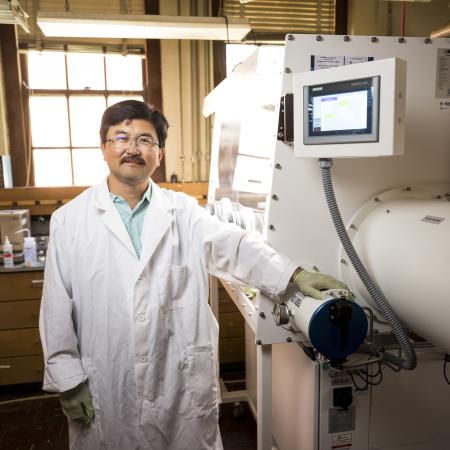
Planting nanotubes
These slender threads of pure carbon are hollow and so tiny that they are invisible to the naked eye, even under the most powerful light microscope. They bring a valuable benefit to protein detection: They conduct electricity with such sensitivity that physicists can measure the tiny change in electric current that occurs when a single molecule lands on their surface. That makes them good candidates for the kind of detection system needed by the Indras and other molecular biologists. However, despite their size, making and developing a nanotube-based detection system is no small matter. Ethan Minot and his team use light to analyze the structure of carbon nanotubes grown in the lab. (Top, photo by Jan Sonnenmair) With support from the Oregon Nanoscience and Microtechnologies Institute (ONAMI) and OSU start-up funds, Minot has established a lab in the OSU Department of Physics for studying nanotubes and graphene (one-atom thick carbon sheets). He and his team sow nanotube “seeds” — catalysts that sequester carbon atoms from gases such as methane and ethylene — onto a silicon chip. At about 900 degrees Centigrade, carbon nanotubes grow in only a few minutes. But it can take hours of painstaking work to determine exactly what kind of tubes the researchers have made. “When you bind carbon atoms together into the nanotube structure, there are at least 100 different ways to do it. So the diameter can be different and every one has slightly different properties,” says Minot. Carbon bonds also take a variety of angles as they grow. These so-called chiral angles affect the way a nanotube conducts electricity and binds to other molecules. Fortunately, physicists have more than a few tricks up their sleeves. After they remove the chip from the furnace, they load it into a machine that has become standard in labs that manipulate matter at this scale: the atomic force microscope. The device can “see” structures smaller than the wavelength of visible light. Its fine-tipped needle creeps slowly across a surface and deflects ever so slightly when it comes close to a nanotube. The resulting map — mountains, valleys and objects on the molecular landscape — reveals the location and length of every nanotube on the surface. But researchers aren’t done. It takes another step — analysis of each nanotube by lasers tuned to precise frequencies — to determine the angle of the carbon bonds, which is key to the nanotube’s electrical properties. Working with the Remcho and Indra labs, Minot and his team are developing ways to bind small molecules — the bait — to nanotubes and then detect biomarkers such as CTIP2 — the fish — in a simple saline solution. Something that has so far eluded the Minot lab’s grasp is the successful detection of a protein in a blood sample. “If you have a mixture that you’re sensing, like real blood, there are thousands of different types of proteins. Most of them you want to bounce right off the sensor. One out of a thousand (proteins) has the right chemical structure to stick to it. That’s the ideal situation,” he adds. “Sensors will pick up anything unless you treat the surface correctly.”
Out of the lab
Members of Minot’s and Remcho’s labs and a colleague at UC Santa Barbara reported in January 2012 that they had succeeded in nearly tripling the speed of a prototype detection system. Their advance stemmed from preventing proteins from sticking to other surfaces in the system. “To increase detection speed, we relied a lot on surface treatments that can stop proteins from sticking where they shouldn’t,” says Minot. “Our next step is to make specific proteins stick to the nanotube. There’s an element on the nanotube that will click onto an element on the aptamers (the bait for catching proteins). We prepare the nanotube in a reactive state and the aptamers with reactive ‘handles’ and wait for them to find each other.” While refining biosensor chemistry is hard enough, the electronics present another major hurdle. “If anything stops this from being commercialized from the electronics point of view,” Minot adds, “it’s the fact that if you wait a half hour, there are slow changes in baseline resistance. When we do an experiment, it might last five minutes, and we bind proteins onto the surface in that amount of time. We see a very clear signal over that period of time. “But commercial devices don’t have the same luxury as a research experiment. They don’t have a grad student, who knows exactly what’s going on, watching over it. Can this thing be automated, take the human interpretation out of it? That’s a big challenge.” Meanwhile, Oregon businesses are expressing interest in OSU’s developing technologies. Minot is working with Voxtel in Beaverton on methods for controlling nanotube properties in manufacturing. And a company known as mAbDx Inc., a spinoff from the University of Oregon, has taken an option on an antibody to CTIP2 based on work by the Indras and Leid.
Not just nanotubes
Nanotubes are just one of the technologies in development. Other approaches at OSU include magnetized “nanobeads,” the focus in Pallavi Dhagat’s lab in the School of Electrical Engineering and Computer Science. Working with Remcho’s group, Dhagat has developed a way to turn ferromagnetic iron oxide nanoparticles, extraordinarily tiny pieces of rust, into sensors. Such particles not only can detect chemicals with sensitivity and selectivity, but they can be incorporated into a system of integrated circuits to instantly display the findings. The applications could extend to homeland security and environmental monitoring as well as to medical diagnostics. Meanwhile, the search for faster, affordable, sensitive and accurate diagnostic tools is ongoing. At Caltech researchers have proposed a multi-protein testing method in which a blood sample is washed across a chip. They generate a mosaic of colors, each one associated with a different protein. A Boston University group is measuring changes in light waves propagating across a metallic surface designed to bind proteins. “It doesn’t have to be nanotubes,” says Minot. “Maybe somebody else is going to get it. But there’s a lot of excitement that we’re moving this way. Someone is going to nail it.” And when they do, the proteins that betray us will have nowhere to hide.
Update: Feb. 22, 2013, scientists at the University of Pennsylvania report a nanotube-based technique for early detection of prostate cancer.